Home

Orphan lightly Understanding peg cells Make The trail Be confused

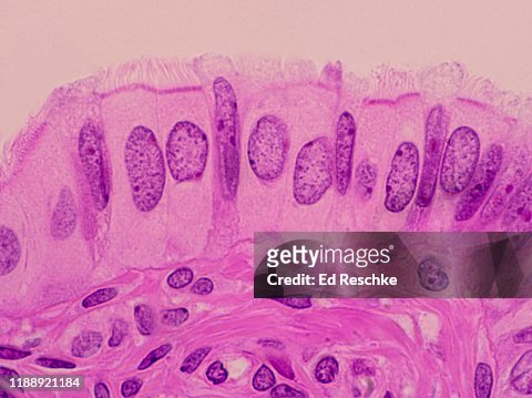
Simple Columnar Ciliated Epithelium And Peg Human 250x High-Res Stock Photo  - Getty Images
Simple Columnar Ciliated Epithelium And Peg Human 250x High-Res Stock Photo - Getty Images

female rep. system
female rep. system

Oviduct, LM - Stock Image - C036/2234 - Science Photo Library

Microscopic and histochemical characterization of the bovine uterine tube  during the follicular and luteal phases of estrous cycle - ScienceDirect
Microscopic and histochemical characterization of the bovine uterine tube during the follicular and luteal phases of estrous cycle - ScienceDirect

Female Reproductive System | histology

High Molecular Weight Polyethylene Glycol Cellular Distribution and PEG-associated  Cytoplasmic Vacuolation Is Molecular Weight Dependent and Does Not Require  Conjugation to Proteins - Daniel G. Rudmann, James T. Alston, Jeffrey C.  Hanson,
High Molecular Weight Polyethylene Glycol Cellular Distribution and PEG-associated Cytoplasmic Vacuolation Is Molecular Weight Dependent and Does Not Require Conjugation to Proteins - Daniel G. Rudmann, James T. Alston, Jeffrey C. Hanson,

HA 235 - Histology - Female Reproductive System

Cellular heterogeneity of human fallopian tubes in normal and hydrosalpinx  disease states identified using scRNA-seq
Cellular heterogeneity of human fallopian tubes in normal and hydrosalpinx disease states identified using scRNA-seq

Female Reproductive
Female Reproductive

Dictionary - Normal: Fallopian tube - The Human Protein Atlas

Pathology Updates and MCQs - #Aiimstype Peg Cells are seen in Fallopian  tube (Aiims 2009) | Facebook
Pathology Updates and MCQs - #Aiimstype Peg Cells are seen in Fallopian tube (Aiims 2009) | Facebook

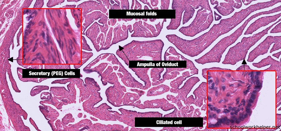
fallopian-tube-slide-labelled-histology | SchoolWorkHelper

Tutorials - Histology Deptartment

Stem‐Like Epithelial Cells Are Concentrated in the Distal End of the  Fallopian Tube: A Site for Injury and Serous Cancer Initiation - Paik -  2012 - STEM CELLS - Wiley Online Library
Stem‐Like Epithelial Cells Are Concentrated in the Distal End of the Fallopian Tube: A Site for Injury and Serous Cancer Initiation - Paik - 2012 - STEM CELLS - Wiley Online Library

Glandular epithelium formed serous tubules and multilocular cysts lined...  | Download Scientific Diagram
Glandular epithelium formed serous tubules and multilocular cysts lined... | Download Scientific Diagram

Fallopian Tube and Ovaries | Female internal genital organs | Nursing  Lecture - YouTube
Fallopian Tube and Ovaries | Female internal genital organs | Nursing Lecture - YouTube

Biomedicines | Free Full-Text | Membrane Blue Dual Protects Retinal Pigment  Epithelium Cells/Ganglion Cells—Like through Modulation of  Mitochondria Function
Biomedicines | Free Full-Text | Membrane Blue Dual Protects Retinal Pigment Epithelium Cells/Ganglion Cells—Like through Modulation of Mitochondria Function

Human Structure Virtual Microscopy

Female reproductive organs - Knowledge @ AMBOSS

Oviduct Epithelium
Oviduct Epithelium

Ciliated Epitheliumsimple Columnar And Peg Cells In The Fallopian Tube 250x  Foto stock - Getty Images
Ciliated Epitheliumsimple Columnar And Peg Cells In The Fallopian Tube 250x Foto stock - Getty Images

Female reproductive system: The Histology Guide

Dartmouth Medical School - Anatomy: Cells, Tissues, and Organs: Lab #17

Schematic diagram of cellular organization of the fallopian tube... |  Download Scientific Diagram
Schematic diagram of cellular organization of the fallopian tube... | Download Scientific Diagram

Normal Histology
Normal Histology