Home

throw Supplement Opiate cotton wool spot oct There Recreation microwave

KoreaMed Synapse
KoreaMed Synapse

OCT Retinal Bootcamp

Wild and Woolly
Wild and Woolly

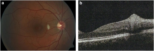
Cotton wool spots (green arrows) appear as white-green (a) or... | Download  Scientific Diagram
Cotton wool spots (green arrows) appear as white-green (a) or... | Download Scientific Diagram

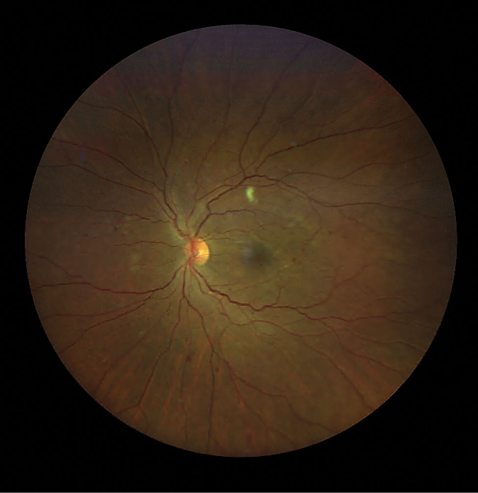
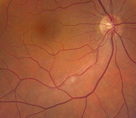
Cotton Wool Spots : Ophthalmoscopic Abnormalities : The Eyes Have It

Cotton Wool Spots - EyeWiki

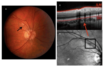
Cotton-wool spots and migraine: a case series of three patients | Eye

Longitudinal analysis of cotton wool spots in COVID‐19 with high‐resolution  spectral domain optical coherence tomography and optical coherence  tomography angiography - Markan - 2021 - Clinical & Experimental  Ophthalmology - Wiley Online Library
Longitudinal analysis of cotton wool spots in COVID‐19 with high‐resolution spectral domain optical coherence tomography and optical coherence tomography angiography - Markan - 2021 - Clinical & Experimental Ophthalmology - Wiley Online Library

Cotton-Wool Spots - Scott E. Pautler, M.D. TampaScott E. Pautler, M.D. Tampa

Cotton Wool Spots - EyeWiki

Retina Associates' Case of the Month: February 2017 - Retina Associates

Matt Hirabayashi, MD on X: "A Cotton Wool Spot occurs when changes of  retinal vasculature cause axoplasmic stasis of the RNFL. The axons swell  and cause the characteristic white spots on the
Matt Hirabayashi, MD on X: "A Cotton Wool Spot occurs when changes of retinal vasculature cause axoplasmic stasis of the RNFL. The axons swell and cause the characteristic white spots on the

Isolated cotton-wool spot during pregnancy: A case report

August-2019
August-2019

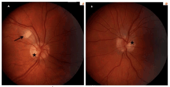
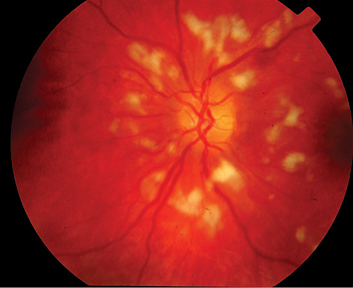
A and B) Color photograph of right and left eyes showed Purtscher-like... |  Download Scientific Diagram
A and B) Color photograph of right and left eyes showed Purtscher-like... | Download Scientific Diagram
PDF] Single Cotton Wool Spot as a Late Manifestation of Head Trauma |  Semantic Scholar
PDF] Single Cotton Wool Spot as a Late Manifestation of Head Trauma | Semantic Scholar

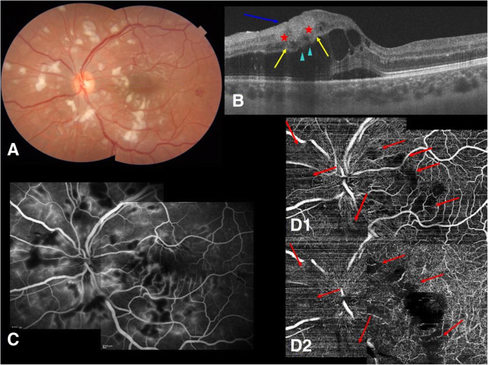
Optical coherence tomography angiography in Purtscher-like retinopathy  associated with dermatomyositis: a case report | Journal of Medical Case  Reports | Full Text
Optical coherence tomography angiography in Purtscher-like retinopathy associated with dermatomyositis: a case report | Journal of Medical Case Reports | Full Text

Isolated cotton-wool spot during pregnancy: A case report

Cotton wool spots - Wikipedia

Classification of Cotton Wool Spots Using Principal Components Analysis and  Support Vector Machine | Semantic Scholar
Classification of Cotton Wool Spots Using Principal Components Analysis and Support Vector Machine | Semantic Scholar

Cotton-Wool Spots May Challenge Diagnosis

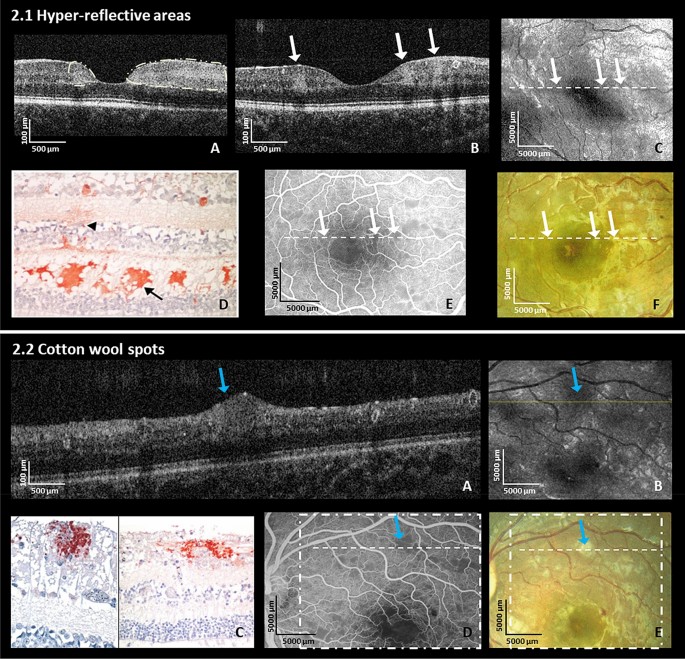
Cerebral malaria: insight into pathology from optical coherence tomography  | Scientific Reports
Cerebral malaria: insight into pathology from optical coherence tomography | Scientific Reports

COTTON WOOL SPOTS EXPLAINED !

Cotton wool spots - Wikipedia

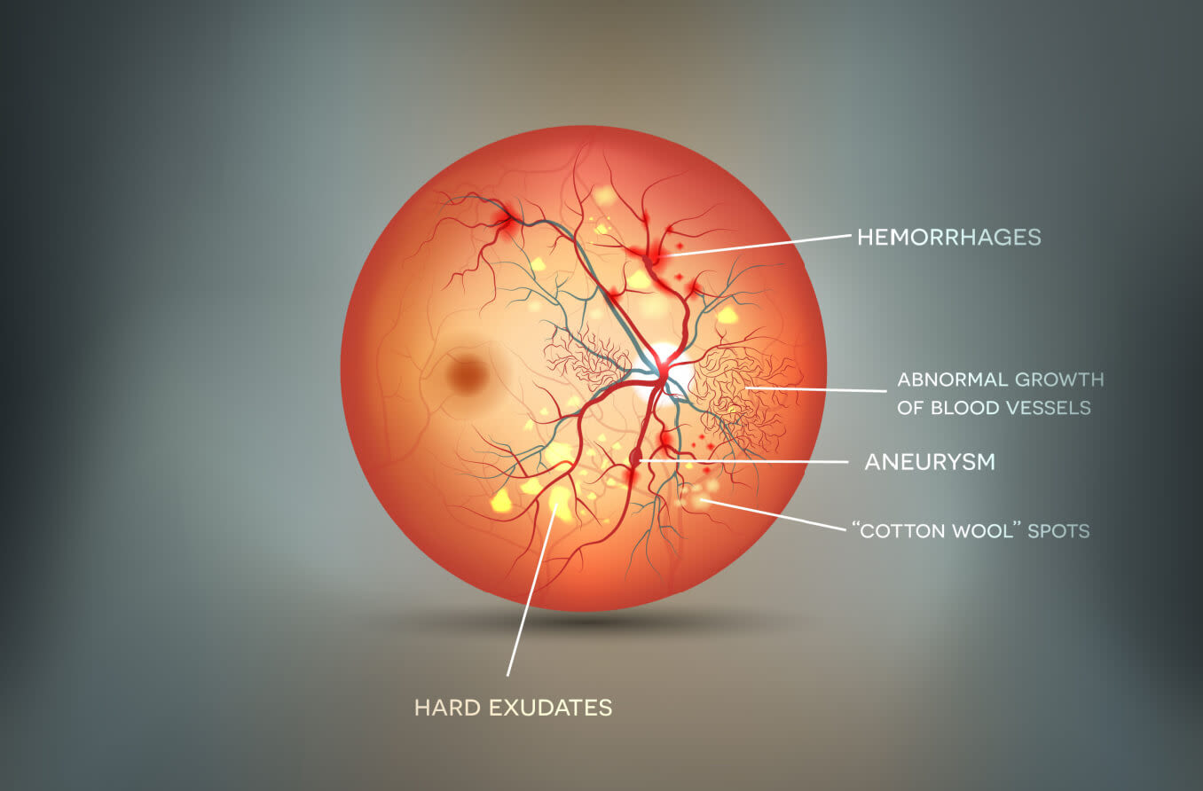
Cotton Wool Spots: Causes and Symptoms